


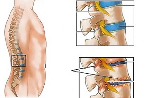
Lumbar osteochondrosis is a chronic disease that develops as a result of a degenerative-dystrophic process in the intervertebral discs. The disease is widespread and affects most people between 25 and 40 years old.
According to statistics, every second adult experiences back pain at least once in his life, while in 95% of cases it is caused by osteochondrosis of the spine.
Patients with a severe course of lumbar osteochondrosis, with persistent pain and other manifestations are known to have a temporary disability. If within four months their condition does not improve, the issue of creating a disability group is decided.
Lumbar osteochondrosis is a serious medical and social problem, as the disease mainly affects people of working age, and in addition, in the absence of treatment, can cause the formation of a disc herniation.
Causes and risk factors
Factors that predispose to the development of lumbar osteochondrosis are:
- spinal structure abnormalities;
- lumbarization - congenital pathology of the spine, characterized by the separation of the first vertebra from the sacrum and its transformation into the sixth (additional);
- sacralization is a congenital pathology in which the fifth lumbar vertebra is joined to the sacrum;
- asymmetric arrangement of joint spaces of intervertebral joints;
- pathological narrowing of the spinal canal;
- reflects spondiogenic (somatic and muscular) pain;
- mbipesha;
- sedentary lifestyle;
- prolonged exposure to vibration;
- systematic physical stress;
- smoking.
Prolonged compression of nerve roots that irritate some organs of the abdominal cavity over time leads to deterioration of their functioning.
Instability of the spinal segment is associated with reactive changes in the bodies of adjacent vertebrae, intervertebral joints, and concomitant spondyloarthritis develops. A significant muscle contraction, for example, against the background of physical activity, leads to a displacement of the vertebral bodies and blockage of the nerve roots with the development of radicular syndrome.
Another cause of pain and neurological symptoms in lumbar osteochondrosis may be osteophytes - bone protrusions in processes and vertebral bodies that cause radicular syndrome or compression myelopathy (spinal cord compression).
Forms of the disease
Depending on the structures involved in the pathological process, lumbar osteochondrosis is clinically manifested by the following syndromes:
- reflex- lumbodynia, lumboishalgia, lumbago; develop against the background of reflex overload of the back muscles;
- oppression (spinal, vascular, radicular)- compression of the spinal cord, blood vessels or nerve roots leads to their development. Examples are sciatica, radiculoemia.
Symptoms of lumbar osteochondrosis
In lumbar osteochondrosis, the symptoms are determined by the structures involved in the pathological process.
Lumbago occurs under the influence of hypothermia or physical overload, and sometimes for no apparent reason. The pain appears suddenly and is of a shooting character. Intensifies when you sneeze, cough, turn your body, exercise, sit, stand, walk. In the prone position, the pain sensations are significantly weakened. Sensitivity and reflexes are preserved, the range of motion in the lumbar spine is reduced.
Observe while touching:
- soreness in the lumbar region;
- paravertebral muscle spasm;
- flattening of the lumbar lordosis, which in many cases is combined with scoliosis.
Nerve root tension syndrome in lumbago is negative. When raising a straight leg, patients notice an increase in pain in the lumbar region, rather than their appearance on an elongated lower limb.
Often, with lumbar osteochondrosis, there is a recurrence of pain attacks, which each time become more intense and prolonged.
In lumbodynia, the clinical picture resembles lumbago, but the increase in pain intensity occurs for several days.
With lumboishalgia, patients complain of pain in the lumbar region that radiates to one or both lower extremities. The pain spreads to the buttocks and back of the thigh and never reaches the legs.
Vasomotor disorders are characteristic of lumboishalgia:
- changes in temperature and skin color of the lower extremities;
- feel hot or cold;
- violation of blood circulation.
The development of lumbar compression syndromes is clinically manifested by the following symptoms:
- dermatomal hypalgia;
- shooting pains;
- weakening or complete loss of deep reflexes;
- peripheral paresis.
With compression syndromes, the pain is exacerbated by torso flexion, sneezing, and coughing.
Diagnosis
Diagnosis of lumbar osteochondrosis is performed based on data from the clinical appearance of the disease, laboratory and instrumental research methods.
In blood tests against the background of lumbar osteochondrosis:
- decrease in calcium concentration;
- increases ESR;
- alkaline phosphatase levels increase.
In the diagnosis of lumbar osteochondrosis, X-ray examination of the spine is of great importance.
Prolonged compression of nerve roots that irritate some organs of the abdominal cavity over time leads to deterioration of their functioning.
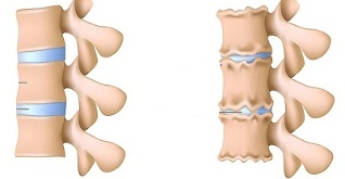
X-ray signs confirming the diagnosis are:
- change the configuration of the affected segment;
- pseudospondilolistema (displacement of adjacent vertebral bodies);
- deformation of closing plates;
- flattening of the intervertebral disc;
- uneven height of the intervertebral disc (a symptom of a spacer), which is associated with asymmetric muscle tone.
Also in the diagnosis of lumbar osteochondrosis, if indicated, the following are used:
- myelography, magnetic resonance imaging or computed tomography - are necessary for persistent symptoms, the development of neurological deficits;
- scintigraphy (study of phosphorus accumulation in the skeletal system, labeled tech-99) - is performed if there is a suspicion of a tumor or infectious process, spinal cord injury.
Differential diagnosis of lumbar osteochondrosis is made with the following diseases:
- spondylolisthesis;
- dyshormonal spondylopathy;
- ankylosing spondylitis (ankylosing spondylitis);
- infectious processes (disc inflammation, spinal osteomyelitis);
- neoplastic processes (primary spinal tumor or its metastatic lesions);
- rheumatoid arthritis;
- deforming osteoarthritis of the hip joint;
- reflected pain (diseases of internal organs and large blood vessels).
Treatment of medial osteochondrosis
For lumbar osteochondrosis, the following treatment tactics are usually followed:
- bed rest for 2-3 days;
- retraction of the affected segment of the spine;
- strengthening the muscles of the back and abdomen (creation of the so-called muscular corset);
- influence on myofascial and myotonic pathological processes.
Lumbago occurs under the influence of hypothermia or physical overload, and sometimes for no apparent reason.
In most cases, conservative treatment of lumbar osteochondrosis is performed, including the following measures:
- muscle infiltration anesthesia with a solution of local anesthetics;
- taking non-steroidal anti-inflammatory drugs;
- taking desensitizing agents;
- vitamin therapy;
- taking sedatives and antidepressants;
- manual therapy, massage;
- physiotherapy exercises;
- acupuncture;
- post-isometric relaxation.
Absolute indications for surgical treatment of lumbar osteochondrosis are:
- acute or subacute pressure on the spinal cord;
- development of cauda equina syndrome, characterized by pelvic organ dysfunction, sensory and movement disorders.
Therapeutic exercises for lumbar osteochondrosis
Physical therapy plays an important role in the complex treatment of lumbar osteochondrosis. Regular exercises allow you to normalize the muscle tone of the paravertebral muscles, improve metabolic processes in the tissues affected by the pathological process and in addition form a well-developed muscular corset that can support the spine in the right position, relieve loadsunnecessary statics by him.
For gymnastics with osteochondrosis of the waist to bring the greatest effect, you must adhere to the following principles:
- regularity of hours;
- gradual increase in the intensity of physical activity;
- avoiding overload during the hour.
Physiotherapy should be performed under the guidance of an experienced instructor, who will select the exercises that are most effective for a particular patient and check the correctness of their application.
According to statistics, every second adult experiences back pain at least once in his life, while in 95% of cases it is caused by osteochondrosis of the spine.
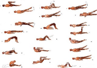
In addition to classes with an instructor, you should perform a series of morning exercises each day, which include specific exercises for lumbar osteochondrosis.
- Relaxation and contraction of the abdominal muscles.The starting position is standing, feet shoulder-width apart, arms sitting on body. Breathe calmly, relaxing the muscles of the anterior abdominal wall. During exhalation, pull the stomach as far as it will go, tensing the abdominal muscles. The exercise should be repeated until slight fatigue appears.
- Head movements with spinal flexion.The starting position is kneeling, leaning on the floor with arms outstretched, back straight. Slowly raise your head and lean on your back. Hold in this position for a few seconds and then return to the normal starting position. Repeat at least 10-12 times.
- "The pendulum".The starting position is lying on your back, arms along your body, legs bent at right angles to the knee and hip joints. Turn your feet left and right in a swinging motion like a pendulum, trying to reach the floor. In this case, the shoulder blades cannot be torn off the floor.
- Boat.Starting position lying on your stomach, arms extended forward. Detach the upper body and legs from the floor, leaning on your back. Hold this position for 5-6 seconds and slowly return to the starting position. Run 10 times.
Possible consequences and complications
The main complications of lumbar osteochondrosis are:
- formation of an intervertebral hernia;
- vegetative-vascular dystonia;
- spondylolysis, spondylolisthesis;
- osteophytosis;
- spondyloarthritis;
- spinal canal stenosis, which leads to spinal cord compression and can cause permanent disability and reduced quality of life.
Prolonged compression of nerve roots that irritate some organs of the abdominal cavity over time leads to deterioration of their functioning. As a result, patients have intestinal dysfunctions (constipation, diarrhea, gas) and pelvic organs (urinary disorders, erectile dysfunction, frigidity, infertility).
Prediction
Pain syndrome in lumbar osteochondrosis occurs in the form of remission and exacerbations. Lumbago lasts 10-15 days, after which the patient's condition improves, the pain subsides. A favorable outcome can be prevented by the associated secondary diseases. Often, with lumbar osteochondrosis, there is a recurrent manifestation of pain attacks, which each time become more intense and prolonged.
Physical therapy plays an important role in the complex treatment of lumbar osteochondrosis.
Patients with a severe course of lumbar osteochondrosis, with persistent pain and other manifestations are known to have temporary disabilities. If within four months their condition does not improve, the issue of creating a disability group is decided.
Prevention
Prevention of the development of spinal osteochondrosis consists of the following measures:
- smoking cessation;
- normalization of body weight;
- improving general physical condition, active lifestyle;
- avoid provocative conditions (weight lifting, sudden movements, turns, bends).